WFU

2017年11月10日 星期五

Edward Chengchuan KO

Edward Chengchuan KO

Head
Division of Oral and Maxillofacial Surgery
Kaohsiung Medical University
Kaohsiung, Taiwan
Takao, Formosa

PhD, The University of Tokyo

Chairman, Membership Committee, 
Asian Association of Oral and Maxillofacial Surgeons
Executive Council, Asian Association of Oral and Maxillofacial Surgeons
Tissue Engineering Lab, College of Dental Medicine, Kaohsiung Med U
Researcher, Dept. of FUJISOFT Cartilage and Bone Regeneration, Tissue Engineering, The U of Tokyo


Most representative Paper for this Takao forum

Edward Chengchuan Ko, Chia-Ming Chang,  Peiying Chang, Chu-Chiang Kao, Kwei-Jing Chen, I-Fan Wu, Michael Yuanchien Chen* Tibial cancellous bone grafting in jaw reconstruction: 10 years of experience in Taiwan. Clinical Implant Dentistry and Related Research 2015;17 Suppl 1:e321-31. SCI類別:DENTISTRY, ORAL SURGERY & MEDICINE 
I.F.= 3.589 (SCI-2014) R/C= 7/88 (8%)


2017年11月9日 星期四

Chang-Wei SU

Dr Chang-Wei SU


Attending Surgeon, DDS
Division of Oral and Maxillofacial Surgery
Kaohsiung Medical University
Kaohsiung, Taiwan
Takao, Formosa


Bachelor of Science, National Taiwan University


DDS, Kaohsiung Medical University


2017年11月8日 星期三

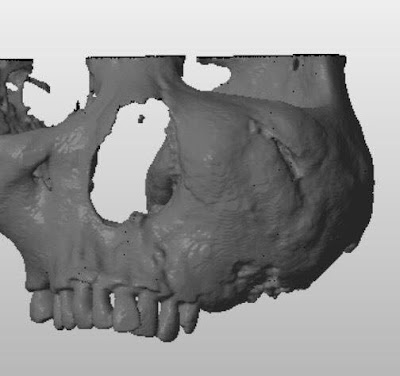
Tumor Excision with Intraoperative Navigation and 3D Printing Technology – A Case Report

Tumor Excision with Intraoperative Navigation and 3D Printing Technology – A Case Report


Chang-Wei SU


Division of Oral and Maxillofacial Surgery
Kaohsiung Medical University
Kaohsiung, TAIWAN
Takao, Formosa


  Great difficulties would be encountered while we would like to excise a deep huge tumor occupying the area in the close proximities to the pterygoid plate, sinus posterior wall and infratemporal fossa. Fatal complications including bleeding and unexpected fracture are of our concern during that kind of surgery. Here we would like to present a case of osteosarcoma with coexistent fibrous dysplasia over the maxilla. Before surgery, we have fabricated a 3D study model in which conveys valuable information and surgical anatomy. Moreover, intraoperative navigation also gave us great assistance in surgical approach as well as precise excision.

Message from President of Asian Association of Oral and Maxillofacial Surgeons

亞洲口腔顎面外科學會理事長致辭
Message from President of Asian Association of Oral & Maxillofacial Surgeons

Message for the 2017 TAKAO Forum of Oral and Maxillofacial Surgery


    On behalf of the Asian Association of Oral & Maxillofacial Surgeons, it is my great pride and honor to extend my warmest greetings and congratulations to the organizers and attendees of the 2017 Takao Forum of Oral and Maxillofacial Surgery.
I witnessed the growing importance and scale TFOMS, which has been successfully held in Kaohsiung Medical University. I highly appreciate the great contribution of all participants in Taiwanese OMS society, either speakers or trainees. Especially Dr Edward CC Ko has built this TFOMS conference in Southern Taiwan to a high-quality international academic conference in the minute of Taiwan OMS society. I would expect the mission of this conference to coincide with the governmental South-Bound policy in the healthcare and academic exchange with many other countries.
Again, I look forward to seeing the success of 2017 TFOMS.


SHOU-YEN KAO, DDS, MHA, DMSc
President, Asian Association of Oral & Maxillofacial Surgeons
Vice-Superintendent, Taipei-Veterans General Hospital
Professor, National Yan-Ming University, School of Dentistry


2017年11月6日 星期一

Recent advances in materials and technology of dental implant

Recent advances in materials and technology of dental implant

Dr Hao-Heung CHANG
Division of Oral and Maxillofacial Surgery
National Taiwan University
TAIWAN


Although the popularity of dental implant with high survival rate has been emphasized in recent years. However, the esthetic problem in anterior teeth region and mechanical failure in posterior teeth have become a new challenge after long-term application. Moreover, facilitating implant inserted in bone with compromised quality and quantity and reducing the healing time also been advocated. It seemed there still a lot clinical unmet of needs should be fulfilled in near future.

In this presentation, experiences and research in developing dental materials and technology in School of Dentistry at National Taiwan University in recent years will be reported. First of all, in order to overcome the drawback in esthetic problem in anterior tooth region, a new zirconia implant has been developed then for marketing. Important findings and results in investigation on such a new zirconia implant from bench to clinical application will be presented. Subsequently, in order to overcome the long-term mechanic failure in dental implant, new abutment with adequate cushion effect mimicked biomechanics of periodontium have been developed and fabricated. Basic mechanics of viscoelasticity of periodontium including creep, stress relaxation and hysteresis and their application in improving implant design will be revealed. Finally, in order to hasten the healing process, different composition of bioceramics have been developed and test in their efficacy in bone regeneration both in vitro and in vivo. A possible roadmap in development of injectable bone cement and three-dimensional printing of composite ceramic materials, based on current results in cell and animal testing will be shown in this presentation.


It is hoped that this presentation will provide a new insight in the new material and technology concerning the dental implant, and provide an opportunity for familiarizing or applying these new materials or techniques soon in the future 

2017年11月5日 星期日

Curvilinear Incision on Iliotibial Tract: Our modified Technique in Harvesting Tibial Cancellous Bone Graft

Curvilinear Incision on Iliotibial Tract: Our modified Technique in Harvesting Tibial Cancellous Bone Graft

Clinical Implant Dentistry and Related Research, Volume 17, Supplement 1, 2015


Edward Chengchuan KO

柯 政全

Head
Division of Oral and Maxillofacial Surgery
Kaohsiung Medical University


Kaohsiung, Taiwan
Takao, Formosa


Background: 

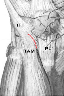
Use of proximal tibia as a donor site has been applied in jawbone reconstruction since the 1990s. Catone and colleagues described a U-shaped incision made on the iliotibial tract during tibial cancellous bone procurement for maxillofacial reconstruction in  1992. We used a curvilinear incision on the iliotibial tract in lateral approach in our tibial
cancellous bone harvesting procedure. 

Objectives: 

The objectives of this retrospective study are to describe our modified lateral approach for procuring cancellous graft from the proximal tibia and to assess the bone volume, donor site morbidity, and associated complications.

Material and Methods: 

Eighty consecutive jawbone reconstructions utilizing autogenous tibial cancellous bone grafts in 78 patients from March 1998 through March 2008 were reviewed. The patient group consisted of 45 males and 33 females, ages 18 to 76 (average age 36.1 1 12.3).Minimal postoperative follow-up period was 3 months. Unlike the traditional U-shaped trapdoor incision on the iliotibial tract, our curvilinear incision was made almost parallel to the fibers of that tract.

Result: 

Only mild complications were observed at donor sites, including temporary paresthesia, gait disturbance, and an unpleasant scar. The average procured graft volume was 17.8 mL. We also present the first case of reconstruction of mandibular continuity defects of up to 6 to 7 cm lengthwise by tibial cancellous bone grafting, which has not previously been reported in the English literature.

Conclusion: 

The modified incision on the iliotibial tract allowed access to obtain an equally good bone volume from the lateral aspect of the proximal tibia, and it rendered wound closure much easier than the procuring techniques described in the earlier literature.


KEY WORDS: bone graft, Gerdy’s tubercle, iliotibial tract, jaw reconstruction, tibia

2017年11月4日 星期六

What Can We Expect in Surgery and Conservative Management for TMD Patients

What Can We Expect in Surgery and Conservative Management for TMD Patients


Shan-Ju Chou D.D.S., MS., FRCD(C), 
Orthodontist and Oral and Maxillofacial Surgeon
Wan-Fang Medical Center
Taipei
TAIWAN


        For people presented with facial pain, difficulty in chewing, and limited mouth opening, many clinical practitioners may prescribe different sort of management , such as medication, occlusal splint, physical therapy, and even though surgery. However, the treatment results are not usually predictable and the practitioners and patients may or may not choose the more invasive intervention if the previous results are not satisfactory. For the treatment accuracy, gathering enough information for exact diagnosis and understanding the pathogenesis of this diseases could direct the treatment to right direction and a better outcome.

        Most commonly, the TMJ patients could be divided into 5 categories: Internal derangement (including degenerative changes), benign tumour, malignancy, traumatic adhesion, neuropathy, and systemic disease. Each category has its own clinical characteristic, and its not difficult to clarify the aetiology by carefully evaluating the pain pattern, disease course, jaw movement and image. Therefore, the more predicable outcome and prognosis are not impossible missions for TMD patients if the adequate treatment performed. 

Computer-aided Mandible Reconstruction: the Beginning Experience

Computer-aided Mandible Reconstruction: the Beginning Experience 


LIU, Yi-Ching


Yunlin Branch
National Taiwan University Hospital


Computer aided system, such as computer-assisted virtual planning, CAD-CAM surgical guides, provides preview of anatomic details in the operation, prediction the cutting line, the inset bone segments in three dimensional pictures and solid models, which make operation easier, safer, and more accurate. Nowadays, computer-aided systems are commonly used in orthognathic surgery, open reduction of traumatic facial bone fractures, and reconstruction after benign tumor resection. These powerful tools also joint oral maxillofacial surgeon, plastic surgeon, prosthodontics, orthodontics, and engineer technicians together, to build a better treatment result for patients.  


Yunlin county has the highest oral cancer prevalence in Taiwan. Patients had their partial mandible resected in conventional surgical methods sometimes suffered from inadequate facial esthetics, malocclusion or mandible side shift. Dentures or dental implant rehabilitation is extremely difficult due to unsatisfied grafted bone position or size. Application of virtual planning and CAD-CAM guide on malignant tumor cases, though, it is still worth a try, to make patients live better after surgery. A case will be presented, which computer-aided mandible resection and reconstruction was performed on an oral cancer patient. With this experience, we defined that flexibility of section margin while tumor progression, reduced preparation time, better adaptation of surgical guides, and better soft tissue management are important future works.

UP to date of MRONJ (medication related osteonecrosis of jaw)

UP to date of MRONJ (medication related osteonecrosis of jaw)

Professor Jae-min SONG


Pusan National University

KOREA

Since Marx reported BRONJ cases in 2003, BRONJ has been reported for more than 10 years, and many studies on the etiology, diagnosis and treatment of BRONJ have been performed in the oral and maxillofacial surgery field. Studies have shown that bisphosphonates have been associated with increased use of Bisphophontate as a treatment for osteoporosis since 1990. In recent years, denosumab has been used as a RANKL inhibitor in addition to bisphosphonates. The jaw necrosis caused by these drugs called MRONJ.
Today I will present about these drugs that cause jaw necrosis, the mechanism, diagnosis and treatment standards of each country. In treatment of MRONJ, Surgical treatment can provide a high level of recovery. We will review the surgical considerations. In addition to surgical treatment, many adjunctive modalities such as LLLT (low level laser therapy), HBO (hyperbaric oxygen therapy), platelet concentrate and fluorescent marking were using.

We will also investigate the recent trends in the use of recombinant hormone, growth factors and stem cells. 

2017年11月3日 星期五

Consideration of temporomandibular joint in occlusion-oriented oral and maxillofacial surgery

Consideration of temporomandibular joint in occlusion-oriented oral and maxillofacial surgery

Professor Tetsu TAKAHASHI


Tohoku University
Sendai
JAPAN


 Temporomandibular joint (TMJ) is a diathrodial joint, which is composed of the temporal bone and the mandible, as well as a specialized dense fibrous structure, the articular disk, several ligaments, and numerous associated muscles. Functionally the TMJ is a compound joint, composed of four articulating surfaces: the articular facets of the temporal
bone and of the mandibular condyle and the superior and inferior surfaces of the articular disk. The articular disk divides the joint into two compartments. The lower compartment permits hinge motion or rotation and hence is termed ginglymoid. The superior compartment permits
sliding (or translatory) movements and is therefore called arthrodial. Hence the TMJ as a whole can be termed ginglymoarthrodial.
 Trauma such as condylar fractures, growth anomalies, neoplastic diseases, and TMJ disorders including osteoarthritis cause dento-maxillo-facial deformities and occlusal disorders. When we correct such dento-maxillo-facial deformities including occlusion, special care should be taken not to aggravate TMJ function such as articulation and occlusion. Therefore, we oral and maxillofacial surgeons should always consider TMJ antomy, pathophysiology, and function. In this lecture, I would like to focus on TMJ, specifically on its biology and function in oral and maxillofacial surgery.


Stability of Simultaneously Placed Dental Implants with Autologous Bone Grafts Harvested from the Iliac Crest or Intraoral Jaw Bone

Stability of Simultaneously Placed Dental Implants with Autologous Bone Grafts Harvested from the Iliac Crest or Intraoral Jaw Bone

Professor Bong-Wook PARK

Department of Dentistry, School of Medicine, Gyeongsang National University, Jinju, Republic of Korea; Head, Department of Oral and Maxillofacial Surgery, 
Changwon Gyeonsang National University Hospital, 
Changwon, Republic of Korea


Background: Jaw bone and iliac bone are the most frequently used autologous bone sources for dental implant placement in patients with atrophic alveolar ridges. However, the comparative long-term stability of these two autologous bone grafts have not yet been investigated. The aim of this study was to compare the stability of simultaneously placed dental implants with autologous bone grafts harvested from either the iliac crest or the intraoral jaw bone for severely atrophic alveolar ridges.
Methods: In total, 36 patients (21 men and 15 women) were selected and a retrospective medical record review was performed. We compared the residual increased bone height of the grafted bone, peri-implantitis incidence, radiological density in newly generated bones (HU values), and implant stability using resonance frequency analysis (ISQ values) between the two autologous bone graft groups.
Results: Both autologous bone graft groups (iliac bone and jaw bone) showed favorable clinical results, with similar long-term implant stability and overall implant survival rates. However, the grafted iliac bone exhibited more prompt vertical loss than the jaw bone, in particular, the largest vertical bone reduction was observed within 6 months after the bone graft. In contrast, the jaw bone graft group exhibited a slower vertical bone resorption rate and a lower incidence of peri-implantitis during long-term follow-up than the iliac bone graft group.
Conclusions: These findings demonstrate that simultaneous dental implantation with the autologous intraoral jaw bone graft method may be reliable for the reconstruction of edentulous atrophic alveolar ridges.

Key words: Simultaneous dental implantation, Severely atrophic alveolar ridge, Autologous bone graft, Iliac bone, Intraoral jaw bone 

Mandibuloplasty- Mandibular Angle Reduciton

Mandibuloplasty- Mandibular Angle Reduciton


Dr Ming-Chih CHANG

Cathay Hospital
TAIWAN

  We will introduce you the indications and procedures of mandibular angle reduction, meanwhile, draw the comparison between the Conventional Angle Ostectomy and the Long-Curved Angle Ostectomy.